
Empanelled with:
ESIC | CGHS | ECHS

Echo Cardiography
Echocardiogram is a test in which ultrasound is used to examine the heart. The equipment is far superior to that used by fishermen. In addition to providing single-dimension images, known as M-mode echo that allows accurate measurement of the heart chambers, the echocardiogram also offers far more sophisticated and advanced imaging. This is known as two- dimensional (2-D) Echo and is capable of displaying a cross-sectional “slice” of the beating heart, including the chambers, valves and the major blood vessels that exit from the left and right ventricle.
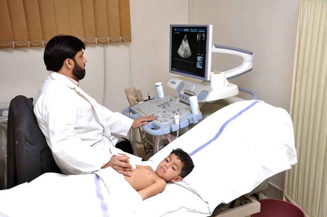
An echocardiogram can be obtained in a physician’s office or in the hospital. For a resting echocardiogram (in contrast to a stress echo) no special preparation is necessary. Clothing from the upper body is removed and covered by a gown or sheet to keep you comfortable and maintain the privacy of females. The patient then lies on an examination table or a hospital bed.
A colorless gel is then applied to the chest and the echo transducer is placed on top of it. The echo technologist then makes recordings from different parts of the chest to obtain several views of the heart. You may be asked to move form your back and to the side. Instructions may also be given for you to breathe slowly or to hold your breath. This helps in obtaining higher quality pictures. The images are constantly viewed on the monitor. It is also recorded on photographic paper and on videotape. The tape offers a permanent record of the examination and is reviewed by the physician prior to completion of the final report.
Echocardiography is an invaluable tool in providing the doctor with important information about the following:
Size of the chambers of the heart, including the dimension or volume of the cavity and the thickness of the walls. The appearance of the walls may also help identify certain types of heart disease that predominantly involve the heart muscle. In patients with long standing hypertension or high blood pressure, the test can determine the thickness and “stiffness” of the LV walls. When the LV pump function is reduced in patients with heart failure, the LV and RV tends to dilate or enlarge. Echocardiography can measure the severity of this enlargement. Serial studies performed on an annual basis can gauge the response of treatment.
Pumping function of the heart can be assessed by echocardiography. One can tell if the pumping power of the heart is normal or reduced to a mild or severe degree. This measure is known as an ejection fraction or EF. A normal EF is around 55 to 65%. Numbers below 45% usually represent some decrease in the pumping strength of the heart, while numbers below 30 to 35% are representative of an important decrease.
Echocardiography can also identify if the heart is pumping poorly due to a condition known as cardiomyopathy (pronounced cardio-myo-puth-e), or if one or more isolated areas have depressed movement (due to prior heart attacks). Thus, echocardiography can assess the pumping ability of each chamber of the heart and also the movement of each visualized wall. The decreased movement, in turn, can be graded from mild to severe. In extreme cases, an area affected by a heart attack may have no movement (akinesia, pronounced a-kine-neez-ya), or may even bulge in the opposite direction (dyskinesia, pronounced dis-kine-neez-ya). The latter is seen in patients with aneurysm (pronounced an-new-riz-um ) of the left ventricle or LV. It must be remembered that LV aneurysm due to an old heart attack does not usually rupture or “burst.”
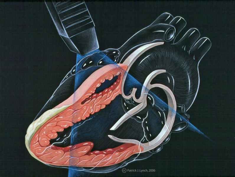
The top diagram on the monitor shows an ultrasound beam (gray triangular area) traveling through the right (RV) and left (LV) ventricle. You can also see the aorta (Ao), left atrium (LA), aortic valve (AV) and mitral valve (MV). Please note that you can review cardiac anatomy and physiology by clicking here. The two pictures on the bottom of the monitor were taken from actual patients. The arrows point to the septum or partition between the RV and LV. The lower left picture demonstrates normal movement of the septum as it moves towards the opposite wall of the LV when the heart contracts. In contrast, the patient on the bottom right has had a heart attack involving the septum. Note that the septum moves sluggishly. Also, it is thinner and “shriveled” as a result of the heart attack.
Valve Function
Echocardiography identifies the structure, thickness and movement of each heart valve. It can help determine if the valve is normal, scarred from an infection or rheumatic fever, thickened, calcified (loaded with calcium), torn, etc. It can also assess the function of prosthetic or artificial heart valves.
The additional use of Doppler helps to identify abnormal leakage across heart valves and determine their severity. Doppler is also very useful in diagnosing the presence and severity of valve stenosis (pronounced stee-no-sis) or narrowing. Remember, unlike echocardiography, Doppler follows the direction and velocity of blood flow rather than the movement of the valve leaflets or components. Thus, reversed blood direction is seen with leakages while increased forward velocity of flow with a characteristic pattern is noted with valve stenosis.
Echocardiography is used to diagnose mitral valve prolapse (MVP), while Doppler identifies whether it is associated with leakage or regurgitation of the mitral valve (MR). The presence of MR frequently prompts the use of antibiotics prior to any dental or non-sterile surgical procedure. Such action helps reduce the rare complication of valve infection.
Volume Status
Low blood pressure can occur in the setting of poor heart function but may also be seen when patient’s have a reduced volume of circulating blood (as seen with dehydration, blood loss, use of diuretics or “water pill.”, etc.). In many cases, the diagnosis can be made on the basis of history, physical examination and blood tests. However, confusion may be caused when patients have a combination of problems. Echocardiography may help clarify the confusion. The inferior vena cava (the major vein that returns blood from the lower half of the body to the right atrium) is distended or increased in size in patients with heart failure and reduced in caliber when the blood volume is reduced.
Other Uses
Patient Preparation
Echocardiography is useful in the diagnosis of fluid in the pericardium (the sac that surrounds the heart). It also determines when the problem is severe and potentially life-threatening. Other diagnoses (plural for diagnosis) made by Doppler or echocardiography include congenital heart diseases, blood clots or tumors within the heart, active infection of the heart valves, abnormal elevation of pressure within the lungs, etc.
Do not come for these investigations immediately after taking meals.

Satyakiran Healthcare is a large medical diagnostic and education conglomerate having national and international presence. Our diagnostic centres are present at multiple locations, presently in five states in the country.
CONTACT US
Satyakiran Healthcare Pvt. Ltd.
Subhash Chowk, Sonipat -131001
Phone : 0130-2242938
: 0130-2242939
: 0130-2242940
: 0130-2242939
: 0130-2242940
Email : [email protected]
Helpline: +91 9812 400 400

Satyakiran Healthcare 2017. Powered by Exact IT Solutions Pvt. Ltd.